Biochemistry Unit
GlycO2Peptide
Core Facilities
The MCBI has outstanding facilities to perform state-of-the-art research. Approaches that are applied range from well validated in vitro and in vivo models with the possibility to validate findings in patient material. Advanced cellular identification and imaging systems are operational (like CYTOF analysis, FACS sorting and analysis, confocal microscopy, life cell imaging) as well as biochemical design and analysis by peptide synthesis and Mass Spectometry. Well characterized in vivo and in vitro models assist in the identification of a number of crucial molecules and processes involved in the development of chronic inflammatory diseases.
The department’s Technology Core Facility comprises an antibody production and Biochemistry Unit (GlycO2Peptide Unit) and the Microscopy and Cytometry Core Facility (MCCF). Scroll down for details on each of them.

Biochemistry Unit
GlycO2Pep
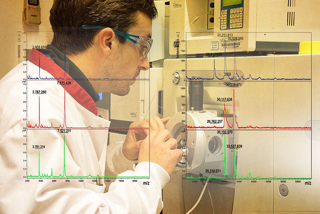
The GlycO2Pep facility is housed at the 12th floor (East Wing) of the O|2 lab building and is managed by Hakan Kalay. GlycO2Pep specializes in two areas, analytical methods for the characterization of glycoconjugates, and the synthesis of peptides and their derivatives. Our facility is equipped with nanoLC-MS systems, an analytical HPLC system, a semipreparative preparative HPLC system and a preparative HPLC system. For peptide synthesis a robotic peptide synthesizer (CEM Liberty) was acquired in 2018. This instrument is able to synthesize peptides in medium/large scale within a day with extremely high levels of purity.
The GlycO2Pep Services
Use the Intern Peptide Ordering Form for:
– peptides synthesis
– peptide derivatives (glycation, lipidation, fluorogenic peptides etc.)
– glycoconjugates
– peptide conjugates
– protein conjugates
– antibody conjugates and labelling
The GlycO2Pep Team
Hakan Kalay (Unit Manager) has expertise in glycomics, proteomics, analytical chemistry, peptide synthesis and bioconjugation techniques. He is also the unit manager and for requests from the unit you can contact him via email h.kalay@vumc.nl.
Juan J. Garcia Vallejo (MD, PhD, Scientific advisor) obtained his PhD in the field of Glycoimmunology and since has a maintained a keen interested in the characterization of glycoconjugates for a better understanding of their function, as well as their synthesis for their exploitation as in biomedicine.
Microscopy Unit
Microscopy and Cytometry Core Facility
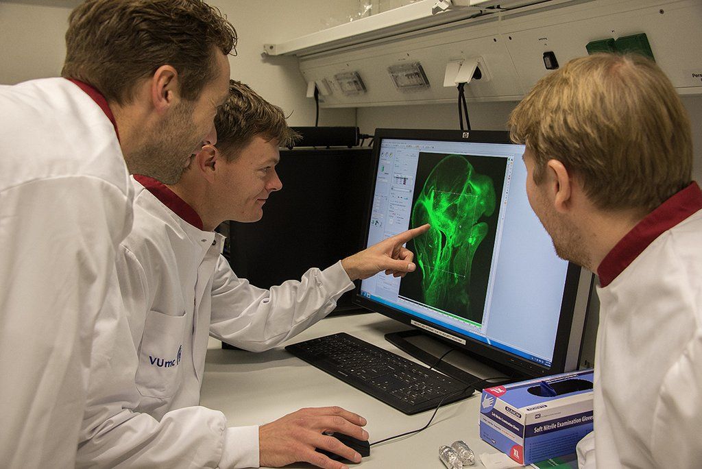
The MCCF (Microcopy and Cytometry Core Faiclity) at Amsterdam UMC – Location VUmc in the O|2 Building is available to all researchers who are included in the Amsterdam UMC network and also to external users and collaborators as a Technology Hotel through the Dutch Techcentre for Life Sciences organization. We provide cutting-edge instrumentation for flow and mass cytometry based immunophenotyping along with high-resolution imaging and high-content screening and other applications and services. Our operators and staff are available to help all users from the early project and experiment planning, to platform selection and work-flow optimization, and onto data analysis and manuscript editing of technical methods.
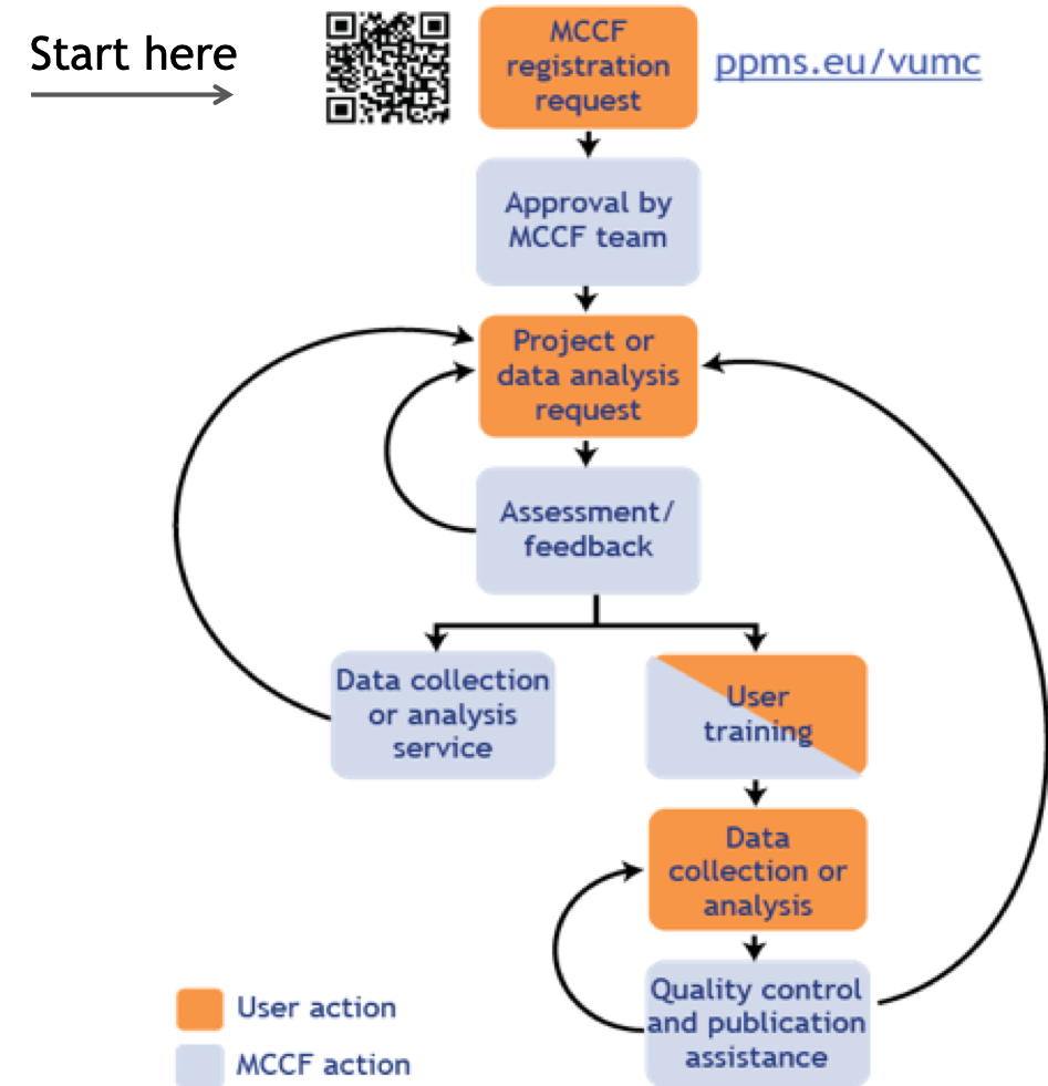
MCCF Workflow
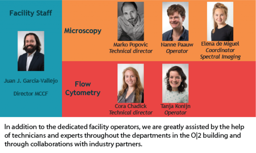
About us
Industry partners
Contract Research
The MCCF provides a full support service for every step in the research process that requires the use of flow cytometry or microscopy techniques:
-Experimental design
-Data acquisition
-Data analysis and quantification
Our team has decades of experience in flow cytometry and microscopy and can assist in the experimental design by advising on appropriate labeling and recording strategies. Our microscopy unit specializes in high-content, live-cell super-resolution, 7+ color multi-spectral imaging and much more.
We routinely use latest data analysis tools for quantification such as FlowJo, FCS Express, NIS Elements, Imaris, Hygens, InForm, QuPath, FIJI, and we are happy to share this expertise with our customers.
In close collaboration with BioBank and VUmc Pathology department we perform a wide array of routine and specialized pathology services: tissue processing, sectioning of paraffin blocks and chromogenic staining.
